Tuesday, December 15, 2015

Novel molecular targets in NASH


NOVEL MOLECULAR TARGETS IN NASH


Non-alcoholic steatohepatits (NASH) is inflammation and damage to the liver due to a build-up of fat.  It is part of a group of conditions referred to as non-alcoholic fatty liver diseases.  It is similar to the kind of liver disease caused by long term, heavy drinking but people with NASH drink little or not at all.


New treatments for inflammatory diseases like NASH could be on the way thanks to a significant discovery made by a group of scientists led by Professor Luke O’Neill.  About eight years ago, they came across the compound MCC950, and began to explore its potential uses. This molecule was previously developed, then abandoned, by Pfizer two decades ago as a possible treatment for arthritis.


These researchers have found that MCC950 directly inhibits NOD-like receptor protein 3 (NLRP3).  NLRP3 is a central mechanism of liver inflammation, injury and fibrosis in various liver diseases including NASH.  The study showed that MCC950 is effective at inhibiting, or reversing inflammation, injury and fibrosis in two different models of NASH. 


This suggests that MCC950 may be used to treat a myriad of inflammatory diseases.  Furthermore, the anti-inflammatory effects of MCC950 were not associated with inhibition of other components of the inflammasome complex that are important for control of infection.


Professor O’Neill and his team hope to form a company to further develop and test this compound.  It could be 10-15 years before it could be fully approved for use in more complex diseases like Multiple Sclerosis or Alzheimer’s.


http://onlinelibrary.wiley.com/doi/10.1002/hep.28175/full


Thursday, November 5, 2015

Diabetes and Your Liver


“Our family, Orange is the New Liver, has been touched by both liver disease and diabetes so it’s no coincidence that I decided to write about the subject. I felt that since November is Diabetes Awareness month it would be the perfect time to talk about diabetes and how it affects me.”
 Morgan d'Organ & Plucky the Orange

Health Changes All

At some time in our life, a medical condition will deeply affect us - whether family, friends, or other loved ones. This is the reason we created Orange is the New Liver, why we are so passionate about our work, and why we are always looking to help others understand their health however, and wherever, we can.

A Family Affair

Four years ago, our CEO’s grandson was diagnosed with Type 1 diabetes. As his career as a racecar driver was ready to take off, he was told he would never race again. However, where there is a will, there is a way. With the help, support, and continuing research by his primary sponsor, the Eli Lily Drug Company our very own Ryan Reed is now the driver of the #16 Drive to Stop Diabetes Xfinity Series car. In honor of Ryan, his dedication, and the continuing work by Lily, we decided to write about diabetes and it’s impact on the liver.

Metabolism and Glucose

Diabetes is an autoimmune disorder that affects your metabolism. Metabolism refers to the way your body uses digested food for energy and growth. Most of what we eat is broken down into glucose. Glucose is a form of sugar in your blood and is the principal source of fuel for your body. Foods are digested and the glucose makes its way into your bloodstream. Your cells use the glucose for energy and growth. However, glucose cannot enter your cells without insulin being present. Insulin makes it possible for cells to take in the glucose.

Insulin is a hormone produced in the pancreas. After eating, your pancreas automatically releases the proper amount of insulin to move the glucose present in your blood into your cells. As soon as glucose enters the cells, blood glucose levels drop.

If you have diabetes, the quantity of glucose in your blood is too elevated (hyperglycemia). This is because your body:

1. Does not produce enough insulin;

2. Produces no insulin;

3. Or, you have cells that don’t respond properly to the insulin your pancreas produces.

This results in too much glucose building up in your blood. The excess blood glucose eventually passes out of your body in urine. Therefore, even though your blood has plenty of glucose, your cells are not getting it for their essential energy and growth requirements.

The Liver and Diabetes

Your liver is an insulin-guided organ and its behavior changes depending on the level of insulin in your body. When levels of glucose (and consequently insulin) are high in your blood, the liver responds to the insulin by absorbing glucose. It packages that sugar into bundles called glycogen. These glucose granules fill up liver cells - the liver is like a warehouse for excess glucose.

When glucose levels drop, insulin production falls too. The shortage of insulin in your blood is the signal that your liver needs to liquidate its assets, sending the glucose stored back into your blood to keep your body well fed between meals and overnight.

The liver doles out stored glucose and has the singular ability to make glucose from scratch, a critical function that keeps people alive when food is scarce. In people with diabetes, however, the liver does not process and produce glucose normally, adding to the challenge of blood glucose control.

The liver cannot directly detect blood glucose levels, it only knows what insulin tells it. Consequently, if there is a shortage of insulin or if the liver does not recognize the insulin, your liver will assume that your body needs more glucose, even if blood glucose levels are already elevated. That is why people with diabetes can have sky-high blood glucose even if they have not eaten. The liver is also responsible for the dangerously high blood glucose levels in people with diabetic Ketoacidosis-a condition in which there is such a severe shortage of insulin that the body cannot process glucose as energy. Instead, the body uses fat. Ketones, waste products created when the liver breaks down fat, can be toxic in large quantities.

In Conclusion

YOUR LIVER IS LARGE AND IN CHARGE! It also processes your body’s fat. Once your liver is full of glycogen, it starts turning the glucose it absorbs from your blood into fatty acids, for long-term storage as body fat. The fatty acids and cholesterol are gathered as ‘fatty packages’ and delivered around your body via your blood. Much of the fat ends up stored in fat tissues. All of this extra fat produced and stored by your liver can sometimes lead the liver itself to get fat: A condition known as non-alcoholic fatty liver disease (NAFLD). The majority of people with diabetes also have NAFLD.


Love Your Liver

Because of the links between diabetes and liver disease, people with diabetes and their health care provider have reason to focus on liver health and use tools such as a liver function test to keep an eye on how you are doing. Your doctor is likely to recommend losing weight, eating well, and exercising. These steps will help control blood glucose levels. For these reasons, what you do for you blood sugar is good for you liver too!

Sunburst Oranges and Orange is the New Liver are the proud recipients of the American Liver Foundation’s first ever “LIVER LOVER AWARD”

Monday, October 19, 2015

Celiac Disease


“I know I’m just a liver so why am I talking about an autoimmune disease? Autoimmune diseases and I are closely related. There are so many autoimmune disorders that affect my function and cause damage that could lead to cirrhosis. I feel it’s important to know all that they can about me and my role in your body.” Thanks, Morgan d'Organ


WHAT IT IS:
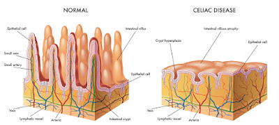
When most of us hear about Celiac Disease, we think of it as a gluten allergy. That is not what it is at all. It is a genetically linked autoimmune disease in which people can’t tolerate gluten. When you eat or use products containing gluten, your immune system sees it as an “invader” and begins a process to attack it.


This attack is aimed at your villi-tiny fingerlike projections on the inner lining of your small intestine. The villi is what absorbs the nutrients and the immune system is trying to keep your body from absorbing the “toxic” substance.

Gluten is a protein found in wheat, rye, barley, and sometimes, oats and occasionally in products like vitamin supplements and even some medications.
SYMPTOMS:
You may experience digestive signs and symptoms, or symptoms in other parts of your body (since it is an autoimmune disorder). Digestive signs are more common in children and can include:
  • Abdominal bloating
  • Chronic diarrhea
  • Constipation
  • Gas
  • Pale, foul-smelling stool or fatty stool (your body can’t absorb fats)
  • Stomach pain
  • Nausea
  • Vomiting
Being unable to absorb nutrients during the years when nutrition is critical to a child’s normal growth and development can lead to other health problems, such as:
  • Failure to thrive in infants
  • Slowed growth and short stature
  • Weight loss
  • Irritability or change in mood
  • Delayed puberty
  • Dental enamel defects of permanent teeth
Adults are less likely to have digestive issues and symptoms but, may instead, have one or more of the following:

  • Anemia 
  • Bone and/or joint pain
  • Canker sores inside your mouth
  • Depression or anxiety
  • Dermatitis herpetiformis – an itchy, blistering skin rash
  • Fatigue
  • Infertility or recurrent miscarriage
  • Missed menstrual periods
  • Seizures
  • Tingling numbness of your hands or feet
  • Weak, brittle bones or osteoporosis
  • Headaches
Intestinal inflammation can cause:
  • Feeling tired for long periods of time
  • Abdominal pain and bloating
  • Ulcers
  • Blockages in your intestine

Celiac Disease can also produce an autoimmune reaction, or a self-directed immune reaction. This can spread outside of your gastrointestinal tract and present in other areas including:
  • Spleen
  • Skin
  • Nervous system
  • Bones
  • Joints
  • Liver

WHO IT AFFECTS:
This disorder can affect both children and adults who are genetically predisposed to it. 1 in 141 Americans have Celiac Disease. It is more common in Caucasians and females and in people with certain genetic diseases.
According to the National Health and Nutrition Examination Survey (NHANES) Study of the CDC, 80% of those with this disorder have yet to be diagnosed.

WHAT IS BEING DONE:
There are currently dozens of clinical trials underway, or concluded, researching every aspect of this disease. At the current time, there is no known cure and the best treatment is a strict adherence to a gluten-free diet. Celiac Disease is a life-long disease managed by lifestyle and generally without medication.

Wednesday, August 19, 2015

MY BABY HAS WHAT?!


My baby has what?!


The initial shock

You hear the words “biliary atresia” and, like most of us, do not know what they mean. Immediately a million thoughts rush through your minds, what is it? What can we do? What medication do we need? Why is this happening to our baby? Then the reality starts to set in as the doctor continues to talk and explain what is wrong with your baby. At first, you do not understand all that is being said- words like biliary system, bile ducts, and Kasai procedure make no sense to you. But then, phrases like no-known-cause and no-known-cure hit you like a ton of bricks. Our precious, seemingly healthy baby but with skin and eyes that are yellowing in color, lovingly stares up at us. In our hearts’ we know this baby is depending on us to help and you realize how helpless you feel. Every fiber of your being is screaming ‘this can’t be real’!

The wait begins

And then the seemingly endless days, then weeks, and sometimes, months in the hospitals have gone by. A continuous parade of doctors, specialists, nurses, and surgeons (the “team”, as they say) politely nod as they pass-by you each day. A new “normal” sets in. You are consumed with the struggle to learn and the search to find out everything you can about biliary atresia in your own battle to save your baby’s life.

You quickly learn

Biliary atresia is a life threatening condition in infants in which the bile ducts, inside or outside of the liver, do not have normal openings. The bile ducts are tubes that carry ‘bile’ from the liver to the gallbladder for storage and to the small intestine for use in digestion. Bile is a yellowish fluid made in the liver that has two main functions:

  1. carry toxins and waste out of your body; and
  2. help the body digest fats and absorb fat-soluble vitamins A, D, E and K.

When the bile duct is blocked, the bile is ‘trapped’ in the liver, rapidly causing damage and scarring of the liver cells (cirrhosis), and eventually liver failure. You know your baby has a tough fight to win!

    Morgan's insight...


    The cause of biliary atresia is not completely understood. It is not contagious and is not passed from mother to child. It is not preventable and not caused by anything the mother did or did not do during pregnancy. Researchers are concluding from studies that the autoimmune mechanisms may be partly responsible. More specifically, the characteristics of the neonatal immune response itself (including Th1 and Th17 responses which are deficiencies in the very important regulatory T-cells), activation of humoral immunity (i.e. a response involving antibodies) or autoimmunity (a “misdirected” immune response that attacks the body itself in error), may explain how and why the unique aspects of the neonatal immune system have gone amiss.

    Symptoms:


    • Jaundice that persists for more than 2 weeks.
    • Dark or brown urine. This is from excessive bilirubin in the bloodstream that passes to the kidneys.
    • Pale or clay-colored (acholic) stools. This is because there is little or no bile reaching the intestines.
    • Enlarged liver that feels harder than normal, enlarged spleen.
    • Poor weight gain.


    Because early diagnosis of this disease is crucial for successful treatment, infants who are jaundiced after 4 weeks of age are usually evaluated for biliary atresia. Your baby’s physician may recommend some or all of the following tests to confirm or rule out this diagnosis.


    • Blood tests. A liver function test (LFT) and blood clotting factors (PT) as well as a complete blood count (CBC); 
    • Liver biopsy; 
    • Ultrasound, to identify any clear abnormalities in the liver or any other organ; 
    • Nuclear scan, which tracks bile flow from the liver; 
    • Sometimes exploratory surgery is employed to examine the liver and bile ducts.

    Once you receive a diagnosis of biliary atresia, a Kasai procedure will take place. This is an operation to re-establish bile flow from the liver to the intestine. During this procedure, the surgeon will remove the damaged ducts outside of the liver and then take a segment of your baby’s own intestine to replace the ducts at the spot where bile is expected to drain. This Y-shaped passageway connects to the liver and to the rest of the intestine.


    Meanwhile


    The wait continues

    You sit, sometimes patiently, sometimes pacing in angst, hoping to hear something, anything, from the surgical team-you can’t believe your sweet baby is having surgery. One day, you had a happy, healthy baby and the next thing you know, you’re sitting in a hospital waiting room…waiting, waiting, waiting.

    When you finally get to see your baby, you are so relieved - but OMG! Tubes are everywhere, equipment is emitting strange sounds and you hardly recognize your baby! Another shocked but experienced parent said, “the more bags hanging and tubes inserted, the longer the stay” and you know this will be a long wait.

    Another “new normal”

    You have learned the Kasai procedure works in about 60% of children and you pray “…please God don’t let our child be in that other 40%!” If the procedure fails, then the term “liver transplant” comes into play and you cannot allow yourself to dwell on that thought!”

    Again, you must adjust to another new normal life. You strain to grasp the new words, like bilirubin, ascites, and NPO, so you may research. The PICU (pediatric intensive care unit) team becomes your extended family-you know everyone’s days off, a bit about their children, their daily schedules, if they are the serious type or use more humor to get through their day. You know where the coffee machine is located, what the cafeteria special will be that day, and who to ask for a pillow or a glass of water. It is your new home!

    Then, hopefully, your baby starts to get better-and with the recovery comes more to know, more to learn and more to do. You are in charge of an array of meds, each to be given at different times on different days. You think… “can I remember all of this and can I administer all of this correctly?” Panic tempers your overwhelming joy but you know you will do it correctly because that is what a parent does! Now you go home and you enjoy your baby crossing each bridge as it comes.

    Knowledge is power!

    Biliary Atresia is one of over 100 diseases that damage the liver, many life threatening and many occurring in infants and young children. Please be aware of those threats to you and your loved ones. Knowledge is power and time is of the essence.

    REFERENCES:

    The Lancet. http://www.thelancet.com/journals/lancet/article/PIIS0140-6736(09)60946-6/abstract
    Cincinnati Children’s Hospital. www.cincinnatichildrens.org/health/b/biliary
    Children’s Hospital of Pittsburgh. www.chp.edu/CHP/atresia
    UCSF. http://pedsurg.ucsf.edu/conditions--procedures/Kasai-procedure.aspx
    U.S. National Library of Medicine. www.ncbi.nlm.nih.gov/pubmed/7472936
    U.S. National Library of Medicine. www.ncbi.nlm.nih.gov/pubmed/ 26090510
    International Scholarly Research Notices. Hindawi Publishing Corp. http://www.hindawi.com/journals/isrn/2012/132089/
    Biliary Atresia Awareness & Research. http://www.biliaryatresia.ca/kasai-procedure/
    Nature Reviews gastroenterology & hepatology. http://www.nature.com/nrgastro/journal/v12/n6/full/nrgastro.2015.74.html

    Thursday, June 4, 2015

    Autoimmune Hepatitis


    “Hey Plucky, would you ever think a liver wouldn’t
    know something about hepatitis?”

    “No way Morgan!”

    “Me either, but I’ve been asked about autoimmune hepatitis and 
    I really don’t know much about it. This is what I know. And, I’ll 
    give you some reference places to find out more”


    AIH in General

    Hepatitis is generally described as injury to the liver with inflammation of the liver cells. Autoimmune Hepatitis (AIH) occurs when your body makes antibodies against the liver tissue because it mistakes your liver as a harmful object and begins to attack it. Although you have injury to your liver, it is more of an autoimmune disease. The exact cause is unknown, AIH, like many autoimmune diseases, is thought to be because of certain bacteria, viruses, drugs, toxins and failure of the immune tolerance mechanisms in a genetically susceptible host. Genetically, the human leukocyte antigens (HLA) on chromosome 6 are the most commonly described association with AIH.


    Chromosome What?

    “Morgan, what in the heck is chromo…whatever 6?”
    Well Plucky, chromosomes are the structures that hold genes. Genes are the individual instructions that tell your body how to develop and function-they govern physical and medical characteristics, such a hair color, blood type, and susceptibility to disease.”
    “I think I’m getting it!”
    “Your body is made up of individual units called cells. You have many different kinds of cells like liver cells, skin cells, blood cells, etc. In the center of most cells you have a nucleus. That is where your chromosomes are located. So, when I said AIH can be due to your genetic susceptibility, I mean there is a genetic abnormality in the chromosome that holds genes related to your immune response-chromosome 6.”

    Potential Causes of AIH

    “OK, back to possible causes of AIH. Viruses such as the hepatitis viruses, measles virus, cytomegalovirus and Epstein-Barr virus can also trigger autoimmune hepatitis. However, this is difficult to assess, as viral infections may occur years before the autoimmune disease manifests symptoms, and the evidence suggesting hepatitis viruses as a trigger are very weak.
    Some medications may be the culprit including oxyphenisatine (a laxative withdrawn by the U.S. Food and Drug Administration), methyldopa, nitrofurantoin, minocycline, atorvastatin, interferon, and diclofenae. 
    Pollutants such as carbon tetrachloride (CCI4), an organic compound once used extensively as a cleaning fluid, refrigerant, and insecticide may also account for the condition.
    Another environmental pollutant is trichloroethylene (TCE), an organic solvent that was widely used as a metal degreaser. 

    Henry Kunkel, 1973


    Discovery and Study

    The earliest writings describe a form of chronic liver disease prevalent among young women and characterized by an excessive increase in serum protein and gamma globulins. In 1951, Kunkel et al, termed the condition “hypergammaglobulinemic chronic hepatitis”. Since then, it has been known by various names including lupoid hepatitis, chronic active hepatitis, chronic aggressive hepatitis, plasma cell hepatitis, and autoimmune active hepatitis.


    Testing and Types

    Today it is simply referred to as AIH and is classified into three types based on serum antibody profiles (blood work). Abnormal values in your liver function test (LFT) will be the first place to check. Type I is the most common with about 75% of people diagnosed while Type II is linked to the other 25%.  There is also Type III which is very rare in the United States.
    Several tests will be ordered to check for various autoantibodies. Type I is characterized by the presence of anti-nuclear antibody (ANA) and anti-smooth muscle antibody (SMA). 25% of patients will have cirrhosis at presentation and have association with other autoimmune diseases such as celiac disease, type I diabetes, rheumatoid arthritis, ulcerative colitis, and autoimmune thyroid disease.
    Type II is characterized by the presence of anti-liver Kidney microsomal (LKM) 1 and/or anti LKM3 and/or anti-liver cytosol 1 (LC1) antibodies. The test for these autoantibodies will usually be a serum protein electrophoresis (SPEP).

    Type III is characterized by the presence of anti-soluble liver antigens/liver-pancreas antibodies (SLA/LP)

    Diagnosis and Treatment 

    Once you have been diagnosed with AIH, the general treatment involves corticosteroids that suppress the immune system. Research indicates most patients achieve full remission in about 3 years but, must stay on the medications for life or they will relapse.
    “As I’ve said before, I’m a liver not a brain and I don’t know much about AIH. This has been a very brief overview and if you would like to learn more, I have included a list of websites that will give you a more in-depth look at autoimmune hepatitis. I also put a list together of some online support groups that might be helpful.”


    REFERENCES
    AASLD Practice Guidelines. Diagnosis and management of Autoimmune Hepatitis.
    Hindawi Publishing Company Journals. Hepatitis research and treatment.
    National Digestive Diseases Information Clearinghouse. National Institute of Diabetes and Digestive and Kidney Diseases. Autoimmune Hepatitis.
    World Journal of Gastroenterology. Autoimmune Hepatitis, one disease with many faces:  Etiopathogenetic, clinico-laboratory and histological characteristics.
    Wikipedia. Chromosome 6 (human).

    ONLINE SUPPORT GROUPS

    AIH Support – provide information and support to people with AIH and their friends and family.
    Drugs.com AIH support group – this is a question and answer forum.
    MD Junction AIH online support group – a community of patients, family members and friends dedicated to dealing with AIH, together.
    I Have Autoimmune Hepatitis – an anonymous support group with information on diagnosis, treatment, symptoms, along with personal stories and experiences with AIH. “You’re not alone.”

    Saturday, March 28, 2015

    Blood Tests Specific to the Liver


    “So Morgan, last time we were talking about getting blood drawn and the Complete Blood Count test. Once I’ve had this, why would I have to have another blood test?”

    “If your CBC comes back with abnormal values Plucky, your doctor will need to know more. You see, the CBC gives you an overview of your health. More specific tests are needed to narrow down where your problem might lie.”

    LIVER FUNCTION TESTS

    Liver Enzyme Tests, formerly called Liver Function Tests, measure various chemicals in your blood made by your liver. Various liver disorders alter the blood level of these chemicals. These tests are not diagnostic of a specific condition; they indicate that there may be a problem with your liver function.

    ENZYME TESTS

    Liver enzyme tests measure current liver cell injury by the amount of enzymes that are leaked out of damaged or dying liver cells. 


    • Alanine aminotransferase (ALT). This is an enzyme that helps process proteins. It is found mainly in your liver cells and to a lesser degree in your kidneys, heart, muscles and pancreas. ALT is measured to see if the liver is damaged or diseased. Low levels are normally found in your blood. When you have a liver disorder, the liver cells spill this enzyme into your blood, raising the level.
    • Aspartate aminotransferase (AST). This enzyme plays a role in your metabolism-the process that converts food to energy. AST is also found mainly in your liver cells and when your liver is damaged the same process happens as with your ALT. However, AST can also be released if heart or skeletal muscle is damaged. For this reason, ALT is usually considered to be more specifically related to liver problems.
    • Alkaline Phosphatase (ALP). This is also an enzyme found mainly in the liver. It can also be found in your bones, intestines, kidneys and other organs. The liver makes more ALP than the other organs or the bones. Some conditions cause large amounts of ALP in your blood. These can include rapid bone growth, bone disease, hyperparathyroidism (a disease that affects how much calcium is in your bones), vitamin D deficiency or damaged liver cells.
    • Lactate dehydrogenase (LD or LDH). This is an enzyme found in almost all of your body’s cells (as well as in bacteria) and is released from cells into the fluid part of your blood (plasma) when the cells are damaged or destroyed. Thus, the blood level of LDH is a general indicator of tissue or cellular damage.
    • Gamma-glutamyl transferase (GGT). High levels of this enzyme are found in the cell membranes in tissues in the liver, bile duct and the kidneys. Bloodstream GGT levels will be higher in people with diseases of the liver and bile ducts. This test can be used to determine the cause of elevated ALP. Both ALP and GGT are elevated in disease of the bile duct and in some liver diseases, but only ALP will be elevated in bone disease. Increased GGT level indicates that a person’s liver is being damaged but does not specifically point to a condition that may be causing the injury. GGT can be used to screen for chronic alcohol abuse (it will be elevated in about 75% of chronic drinkers). 
    • 5 “N” Tase (5’NT). Higher levels of this enzyme will indicate a problem with bile secretion. Hepatitis C or cirrhosis can cause blockage of bile flow.


    PROTEIN TESTS

    Liver function tests look at levels of proteins made by the liver.

    • Bilirubin (BIL). When red blood cells complete their lifecycle and breakdown naturally, they produce bilirubin. It’s a yellow pigment that’s passed on to the liver and excreted in your bile. Most of the time, the body produces as many red blood cells as it breaks down. However, if the red blood cells break down more rapidly or if liver function becomes impaired, bilirubin levels in the blood rise. In patients with Hepatitis, bilirubin levels tend to fluctuate. A prolonged persistent rise in bilirubin for a patient with chronic Hepatitis C usually indicated severe liver dysfunction. When your bilirubin is high the whites of your eyes and your skin will be a yellowish color (jaundice). This is one of the three tests used to determine your wait time for a liver transplant. There are two measurements for your bilirubin.
      1. Total bilirubin (unconjugated). This measures the levels of all the bilirubin in your blood.
      2. Direct bilirubin (conjugated). Measures the bilirubin that has been processed by the liver and attached to other chemicals.
    • Albumin (ALB). As we’ve talked about, is the major blood protein made by the liver. One function is to keep the blood from leaking through your blood vessels, which can cause fluid retention in the ankles (edema), lungs or abdomen (ascites). Low levels of albumin may be due to liver injury or kidney disease, malnutrition or even a low protein diet.
    • Prothrombin time (PT). This measures how quickly your blood clots, which is dependent on clotting factors (proteins) that are made by the liver. PT is used as a marker of advanced liver disease and can indicate blood clotting problems where it takes you longer to stop bleeding. Your lab may also give PT results that have been converted to an internationally recognized and easily comparable value that’s called the International Normalized Ratio (INR). The INR is one of the three factors used to determine wait time for a liver transplant.
    • Alpha-fetoprotein (AFP). This test is used as a marker for potential liver cancer. Not everyone with liver cancer has this marker. Pregnant women usually have higher levels of this protein which is also used to look for problems in pregnancy. You may have higher levels of this protein if you have hepatitis or cirrhosis.
    • Total Protein (T/P). This test measures the two main proteins in your blood: albumin and globulin. This test will give your A/G ratio. Normally there is a little more albumin than globulins, giving a normal A/G ratio of slightly over 1. Because disease affects the relative amount of albumin and globulins, the A/G ratio can provide a clue as to the cause of the change in protein levels. A low A/G ratio may reflect over production of globulins, such as seen in autoimmune diseases, or an underproduction of albumin such as may occur with cirrhosis, or it could be selective loss of albumin from the circulation, as may occur with kidney disease. A high A/G ratio suggests underproduction of immunoglobins as can be seen in some genetic deficiencies and in some types of leukemia.


    “Oh my, look at the time! I’m sorry Morgan but I have to go. This is really great information you’re sharing with me but I have a photo shoot to do. As you know, Sunburst Oranges was the recent recipient of the American Liver Foundation Greater Los Angeles division’s first annual “Liver Lover” award and I am accepting of their behalf.”

    “That is so great Plucky! I have seen you and Sunburst Oranges at some of the Liver Life Walks working really hard to spread awareness about liver disease and sharing those delicious Plucky brand oranges. I’ll finish this up and you can read it later. Just a couple of things before you go. Would you bring me back some of those oranges? And, can I have your autograph?”

    "Very funny Morgan!"

    “O.K., where were we? Yes, we just finished with the blood tests specific to the liver. I’m including one more because it is the third test used to determine your wait time for a liver transplant.”

    OTHER TESTING

    • Creatinine, is a breakdown product of creatine, which is made by the liver and transported to your muscles. Your kidneys excrete the waste product creatinine and, when your kidneys are damaged, creatinine levels rise. When your liver stops functioning in end-stage liver disease, this can cause serious kidney problems as well.

    Lab tests, or bloodwork, can give important clues about your overall health and the health of your liver. I strongly suggest keeping a copy of all lab results. This will give you your “baseline” and allow you to watch for changes in your health over time. This will also help you, and your doctor, to check the impact of any treatments you undergo.


    Again, factors such as age, gender, stress, meds, infections and a host of other things can all affect your test results. So, keep all these factors in mind when reviewing your lab results. Understanding your test results may seem confusing at first. But, they can help you take charge of your health and understand why your doctor prescribes certain tests and medications.

    With practice, over time, it will all become easier to understand. "I've got to go see what our new 'rock star' plucky is doing at this photo shoot so, until next time, LOVE YOUR LIVER!"

    Morgan d' Organ


    Thursday, March 26, 2015

    Complete Blood Count

    COMPLETE BLOOD COUNT

    “Morgan, what do you mean, they stick you with a needle and suck out some of your blood? I don’t think I want to do that!”


    “It really doesn’t hurt Plucky. And blood tests are an easy, non-invasive way of letting your doctor know what is going on inside of you.”

    The first basic blood test, whether you’re having a routine physical or if a problem is suspected, is a Complete Blood Count (CBC). This is not a diagnostic test, it is the starting point for a diagnosis. If this test comes back normal, great, go on your merry way and have a great day. If it shows you have any abnormal values, it tells your doctors where these abnormal values are and a more specific test will be ordered.

    Remember, your blood generally consist of 2 major parts, plasma and cellular elements. The plasma is the liquid part of your blood and allows it to flow easily. The other part consists of blood cells (red blood cells, white blood cells) and platelets. The Complete Blood Count (CBC) measures the cellular part of your blood, plus RBC indices. The RBC indices provide information about your hemoglobin content and size of your red blood cells.


    The following is what your CBC will be measuring:

    White blood Cells (WBC) fight infection. There are many types of WBCs and each fights infection in a special way.

    Neutrophils: An important infection fighting WBC. The number your doctor looks at is your absolute neutrophil count (ANC). A healthy person has between 2,500-6,000.

    Band cells: Slightly immature neutrophils.

    T cells – T type lymphocytes: This WBC is of key importance to the immune system and is at the core of adaptive immunity, the system that tailors the body’s immune response to specific pathogens. They are like soldiers who search out and destroy the targeted invaders. T cell activation is measured as a way to assess the health of patients with HIV/AIDS and less frequently in other disorders.

    B cells – B type lymphocytes: This WBC works chiefly by secreting antibodies into your body’s fluids. Each B cell is programmed to make one specific antibody. For example Plucky, one B cell will make an antibody that blocks a virus that causes the common cold, while another produces an antibody that attacks the bacteria that causes pneumonia. When a B cell encounters the kind of antigen that triggers it to become active, it gives rise to many large cells known as plasma cells, which produce antibodies.

    Monocytes: This type of WBC develops in your bone marrow then goes into your blood, where they circulate for a few days and then go into your tissues. They then develop into one of two mature WBCs. Either dendritic or macrophages.

    Eosinophils: These WBCs become active when you have certain infections from parasites, autoimmune diseases, eczema, hay fever or leukemia. Low levels indicate alcohol intoxication or over production of certain steroids such as cortisol.

    Basophils: These WBCs contain the anticoagulant heparin and also histamine which promotes blood flow to tissues.


    RBC Red Blood Cells carry oxygen to and carbon dioxide away from the cells in your body. The CBC measures RBCs in many ways, but the simplest measure is either Hemoglobin (Hgb)-the part of each RBC that carries iron or Hemocrit (Hct)-the percent of RBCs in the blood. When the Hgb and Hct fall too low, it’s called anemia.

    MCV Mean Corpuscular (cell) Volume measures the average amount of space occupied by each RBC. A high MCV can indicate liver disease and/or alcohol abuse.

    MCH Mean Corpuscular (cell) Hemoglobin measures the average amount of oxygen-carrying Hgb found in your RBCs. A high MCH can indicate anemia. Alcohol abuse can also be a contributing factor of high MCH.

    MCHC Mean Corpuscular (cell) Hemoglobin Concentration. This is an Hbg/Hct ratio. A calculation of the average percentage of hemoglobin inside a RBC.

    I want to add that corpuscular refers to blood cells.


    RDW Red Cell Distribution Width is a measurement of the amount that RBCs vary in size. A high RDW means that your RBCs vary a lot in size. To determine a possible cause for this, a comparison is made to the MCV. One cause can be liver disease, another can be a type of anemia.

    PLT Platelets help control bleeding. The medical term for platelets is thrombocytes. Platelets are produced in your bone marrow. You may bruise easily when your platelet levels are low and you may bleed even when you’re not injured. Your risk of bleeding goes up when your PLT drops below 20,000. A high or low PLT count is a sign of a blood or bone marrow disorder. A low PLT count is seen with viral infections, certain drugs such as acetaminophen, leukemia, autoimmune disorders and various other causes. Additional blood test will be required for this.

    MPV Mean Platelet Value is an indicator as to whether the bone marrow is manufacturing PLTs normally. An MPV measurement can enable your doctor to detect a problem before it shows up in a PLT test.

    NOTE: Your doctor may order a WBC differential test. This would include absolute neutrophil count or % neutrophil, absolute lymphocyte count or % lymphocytes etc… This is to determine the cause of abnormal results of your WBC on your CBC.

    The following is what the CBC portion of your lab results might look like:


    Each lab has different normal ranges and each person’s normal can vary depending on your age, gender, ethnicity and a few other things. Therefore, I did not talk about normal ranges of any test. If you fall out of your normal range, it will be clearly indicated so you will know. Usually with something like an *, an H if it’s high or an L if it’s low.


    “Morgan, why do I need a blood test, I’m not sick or anything?”


    “Well, a blood test tells your doctor if there is a problem before you even feel any symptoms. Early detection, in most cases, will result in a better prognosis. Do you know that I have a dear friend who was feeling fine and had a blood test that came back with a very high WBC count. This could mean she had a really bad infection or maybe even leukemia! Of course, the doctor ordered several other blood tests. All additional tests came back normal thank goodness. The doctor told her abnormally high white blood cell count could have been caused by stress in her life or even an error at the lab. Anyone can make a mistake. But, the most important thing to remember is “knowledge is power”. And, the sooner you have information about a health crisis, the better the outcome will be.”

    Morgan d Organ

    Thursday, January 29, 2015

    Liver Awareness - Globulins

    “Morgan here checking in with you again as we are finally done celebrating the New Year”.

    “To refresh your memory Plucky, we were talking about the proteins your liver produces. The last time we were discussing Albumin. Now we are going to dive into Globulins.”

    I am telling you about these things because I looked at a blood panel report one day and could not understand much of it. All the symbols and numbers were so intimidating and confusing! It is scary being sick and not knowing what is going on inside you! If you learn a little more about yourself, you will be better equipped to understand some of the things your doctor discusses and maybe even know what is in those confusing reports. “


    C’mon Plucky, time to 
    get back to work!


    “Today I’m going to explain Globulins and how they affect your health. Next time we’ll try to unravel those blood panel test results.”

    GLOBULINS

    As we have talked about before, proteins are the most abundant compounds in your serum (the rest of your blood after the red blood cells, white blood cells and platelets have been removed). Amino acids are the building blocks of all proteins. In turn, proteins are the building blocks of all cells and tissue. They are the basic components of enzymes, many hormones, antibodies and blood clotting agents. Proteins act as a transport substance for hormones, vitamins, minerals, lipids and other material. Lipids are fat-like substances in your blood and tissues. Your body needs a small amount of lipids to work normally.


    Proteins also help balance the osmotic pressure of your blood and tissue. Osmotic pressure is part of what keeps water inside a particular compartment of your body. Other proteins play a major role in maintaining the delicate acid-alkaline balance of your blood (pH). pH stands for potential hydrogen. In addition, serum proteins serve as a reserve source of energy for your tissues and muscle when you are not ingesting enough.

    “Good grief Morgan, what does all that mean?”

    “First Plucky, your blood tells a lot about you and even though it sounds complicated you need to know a few terms. Second, Plucky, your osmotic pressure has to do with the pressure on the outside of your capillaries while your blood pressure is pressure from inside your capillaries. This is important because this is how your blood keeps at correct density and smoothly flows through your veins and arteries. When your Albumin level is low (osmotic pressure is low), it allows too much water into your cells causing edema. You know, like when your ankles or feet swell.  Now back to the Globulins:

    Chemical structure of human serum
    albumin (HSA). HSA is the most
    abundant protein in blood plasma
    and is an important transport protein.
    The major serum proteins are divided into two groups: 
    Albumin and Globulins. Globulins are a group of proteins in your bloodstream that help to regulate the function of the circulatory system. (Your circulatory system is responsible for transporting materials throughout your entire body.  It transports nutrients, water and oxygen to your billions of body cells and carries away wastes such as carbon dioxide that body cells produce.) Globulins are produced in the liver and immune system. They have multiple different functions; the group includes immunoglobins, enzymes, carrier proteins and complement. Your globulin levels will affect the amount of ample proteins in your bloodstream. If these proteins are not kept at the proper level, it can be difficult for your body to fight infections, clot, or transport nutrients to the muscles.

    Human Circulatory System
    Globulin is made up of different proteins called alpha, beta and gamma. The alpha and beta globulins are made by the liver, while the gamma globulins are made by the immune system. Certain globulins bind with hemoglobin, hemoglobin is another protein in your blood that carries oxygen from your lungs to your tissue and returns carbon dioxide back to your lungs. Other globulins transport metals, such as iron in the blood that help fight infection.

    At certain times when your Doctor needs to get more information from your blood, serum globulin can be separated into several subgroups by Serum Protein Electrophoresis Test (we will go into the details of why and how later).


    SUBGROUPS OF GLOBULINS

    Lipoproteins of the blood, LDL and
    HDL (cholesterol)

    • Alpha 1 globulin (α1) – This fraction consists of:
    High-density lipoprotein (HDL) the good type of cholesterol. Transcortin, aka corticosteroid binding globulin (CBG) or (serpin A6). Serpin is a serine protease inhibitor. Transcortin is a major transport protein for glucocorticoids and progestins. These are the hormone transporters I was telling you about, more specifically, estrogen. A female hormone.

    α1 antitrypsin (α1AT) a protease inhibitor. This is the major protein associated with alpha 1 globulins. It inhibits a wide variety of proteases. Protease, also called proteinase or peptidase, is a type of enzyme that occurs naturally in living things and forms part of many metabolic processes. They form part of the larger systems in the body, including the digestive, immune, and blood circulation systems. When you have a virus (HCV or HIV) in your body, the virus uses a healthy cell for replication. It does this by making the healthy cell produce certain proteins the virus can use to make more copies of itself. Two of the proteins used by the virus are reverse transcriptase and protease.


    “Time-Out Morgan! I’m lost again. 
    What’s does that mean to me?

    “OK Plucky, that was a mouthful, I agree. However, it has to do with your cholesterol and I know you know about that. The protease inhibitors are important when your body is helping me fight viral infections like HCV and HIV. The HCV virus survives by replicating itself at a high rate. It does this by making healthy cells produce proteins that the virus needs to survive. Your healthy cells will produce proteases. You’ve heard about the new protease inhibitor drugs. These assist the protease inhibitors you already have!


    • Alpha 2 globulin (α2) - Consists of:

    Haptoglobin (HP). Binds the globin portion of free hemoglobin that’s made when red blood cells die (red blood cells have a life of 110-120 days). Haptoglobin transports this back to the liver for recycling. Haptoglobin is known as an acute phase reactant. Its level increases during acute conditions such as infection, injury, tissue destruction, some cancers, burns, surgery or trauma. Its purpose is to remove damaged cells and debris and rescue important minerals such as iron.

    Macroglobulin (α2M). A protease inhibitor and one of the largest plasma proteins. It transports hormones and enzymes, exhibits effector and inhibitor functions in the development of the lymphatic system and inhibits components of the complement system and hemostasis system. Meaning, they act as back up inhibitors when inhibitors that are more specific are consumed during traumas which lead to major activation of the proteolytic cascade in coagulation and fibrinolysis. A proteolytic cascade occurs when several proteases activate each other; and, Fibrinolysis is a process that prevents blood clots from growing and becoming problematic.

    Prothrombin (Pro). A coagulation factor that is needed for normal blood clotting. Prothrombin is a precursor to thrombin. Tests will measure prothrombin time (PT).

    Ceruloplasmin (CER). Transports copper. An acute phase reactant. It is frequently elevated when you have inflammation, severe infection, tissue damage, and may be increased in some cancers. It is reduced in Wilson’s Disease.

    • Beta globulins (β) – Consists of:
    Low-density lipoproteins (LDL). The bad type of cholesterol.

    Transferrin (TF). The main protein in the blood that binds to iron and transports it throughout the body. Beta globulins consist mainly of transferrin. The amount of transferrin that is available to bind to and transport iron is reflected in measurements of the total iron binding capacity (TIBC), unsaturated iron binding capacity (UIBC), or transferrin saturation.

    Microglobulin (β2m). A class 1 antigen is a marker that is increased in serum during immune activation. Β2m values have emerged as markers for the activation of the cellular immune system, as well as a tumor marker in certain hematologic malignancies. Urine β2m values in both serum and urine can help distinguish a problem of cellular activation from a renal disorder.

    “So Morgan! I think I am starting to get it! My blood is the lifeline of all those things my organs need to keep functioning properly, keep my immune system strong to fight invaders in my body. And help distribute all those proteins I need. WOW. Plus a sample of my blood will let the Doc know where something is going wrong in my body. Is that right Morgan?"

    You’re getting the hang of it Plucky. The body is very complex and your blood provides you with nutrients and hormones that must get to the right destination. Healthy blood is very important for your life. Now we can finish up with the Gamma Globulins as they are important to your immune system:

    • Gamma globulins (γ)
    These proteins are also called antibodies. These globulins are the only globulins not produced in the liver; they are produced by the immune system. They help prevent and fight foreign substances (antigens), such as bacteria or viruses, causing them to be destroyed by the immune system.  

    The most significant gamma globulins are immunoglobulins (Ig). Your body makes different immunoglobulins to combat different antigens. Sometimes your body may even mistakenly make antibodies against itself, treating healthy organs and tissues like foreign invaders – this is called an autoimmune disease.

    THERE ARE 5 SUBCLASSES OF ANTIBODIES:

    Immunoglobulin A (IgA). Found in high concentrations in the mucous membranes, particularly those lining the respiratory passages and gastrointestinal tract, as well as saliva and tears.

    Immunoglobulin G (IgG). The most abundant type of antibody. It’s found in all body fluids and protects against bacterial and viral infections.

    Immunoglobulin M (IgM). Found mainly in the blood and lymph fluid. It is the first antibody to be made by your body to fight a new infection.

    Immunoglobulin E (IgE). Associated mainly with allergic reactions.

    Immunoglobulin D (IgD). This exists in small amounts in the blood and is the least understood antibody.

    IgA, IgG and IgM are often measured together to give your doctor important information about your immune system function, especially relating to infection or autoimmune disease.

    “As you can see, your blood protein levels can tell your doctor a lot about what is going on with your body. Blood tests are routinely performed and I want you to be familiar with some of the symbols so as to better understand them. I’m going to talk about blood tests and how to interpret them next time. I’ll also go into what the normal ranges should be. Although normal values do vary from lab to lab. So, until next time, enjoy life and love your liver!”


    Morgan d’Organ